Radiological reports. Explained simply. By an expert.
Get a picture of your symptoms yourself and thus get the key to successful therapy & freedom from painMRI second opinion online. Understand MRI findings – Have MRI findings explained.
Make an appointment

Quick and easy online appointment booking.
Transfer images

Digital transfer of the radiological images you wish to consult.
Personal Consultation

Your personal consultation with Dr. Friedrich via video call.

Associate Professor Dr. Friedrich, PhD
Medical University of Vienna
Dr. Friedrich, PhD
Associate Professor of Radiology
“A picture is worth a thousand words. That’s why I want to help patients to get a picture of the cause of their pain themselves.
Like an independent interpreter, I translate the content of the MR images and findings for the patients so that they are well prepared to play an active role in therapy planning with their attending physician.
This makes a lot of sense, as we know from numerous studies how important “compliance”, i.e. the patient’s “commitment”, is for the success of the therapy.”

Austria’s top specialist for MRI of the joints & spine: “Instructor” of the German Society for Musculoskeletal Radiology for Austria.

More than 20 years of research & teaching in the field of MRI joints and spine:
Medical University of Vienna
,
New York University,
University of Ottawa.
Head of the Musculoskeletal Radiology working group of the Austrian Radiological Society for many years. Senior consultant at the Department of Radiology at the Medical University of Vienna.
Founder of Austria’s first private MRI practice specializing in the area of joints and the spine.
Initiator of one of the most successful continuing education programs at Europe’s largest radiology congress, the European Congress of Radiology (ECR): Case-Based Diagnosis Training.
More than 20 years of research & teaching in the field of MRI joints and spine:
Medical University of Vienna
,
New York University,
University of Ottawa.
Austria’s top specialist for MRI of the joints & spine: “Instructor” of the German Society for Musculoskeletal Radiology for Austria.
Head of the Musculoskeletal Radiology working group of the Austrian Radiological Society for many years. Senior consultant at the Department of Radiology at the Medical University of Vienna.
Founder of Austria’s first private MRI practice specializing in the area of joints and the spine.
Initiator of one of the most successful training programs at Europe’s largest radiology congress, the European Congress of Radiology (ECR): Case-Based Diagnosis Training.
Numerous national and international lectures, book contributions, publications in specialist journals.

Map of the instructors of the German Society for Musculoskeletal Radiology (DGMSR)
The digital transfer of your radiological images / MRI images is at your own risk.
You can use a file hosting service of your choice for this. A free and easy-to-use example is
www.swisstransfer.com.
For “Send file to” or “Email to” please use the following email address:
mrt@talk2friedrich.com
Please send the entire contents of the CD that you received from your radiologist or at the hospital.
Please note that the images must be sent at least 2 hours before your personal consultation with PD Dr. Friedrich.
Your conversation with Dr. Friedrich, PhD, will take place via “Zoom” ( video call).
The conversation consists of 3 parts:
1. You describe your complaints or show where you have pain, how long these complaints have existed, whether there was a trigger for them and how they manifest themselves.
2. Dr. Friedrich, PhD, explains and shows you what can be seen on your radiological images in a way that is easy to understand.
3. Dr. Friedrich, PhD, will discuss with you how likely it is that the changes in your images are responsible for your symptoms and what options are available for further action. This will enable you to form your own picture of your situation and, together with your treating physicians and therapists, start the right therapy for you with motivation.
Before the appointment for your personal consultation with Dr. Friedrich please make sure
that you have successfully installed the
“Zoom”
app on your cell phone or computer,
that you have carried out all updates to the program,
that you have activated the “End-to-end encryption” option in the Zoom settings and
that you are finally logged in / registered in Zoom in good time before your appointment.

Have MR images explained - independently & professionally
Many patients receive MRI results that are difficult to understand. As an independent specialist, I will help you to better understand your MRI results – without any vested interest in one or the other therapy option.
How I assess your MR images
You send me your MR images. I will analyze them carefully and give you an easy-to-understand assessment in a personal consultation via video call.
Not every change in the MRI requires therapy – I will show you what is relevant and what is not.
Why ambiguities are so common
Translating MRI findings - understandable for laypeople
Terms such as “chondrosis”, ‘protrusion’ or “degenerative” can be unsettling or even frightening. Let me explain what really lies behind them.
Using concrete examples, I explain typical MRI findings and what they mean – realistically and objectively.
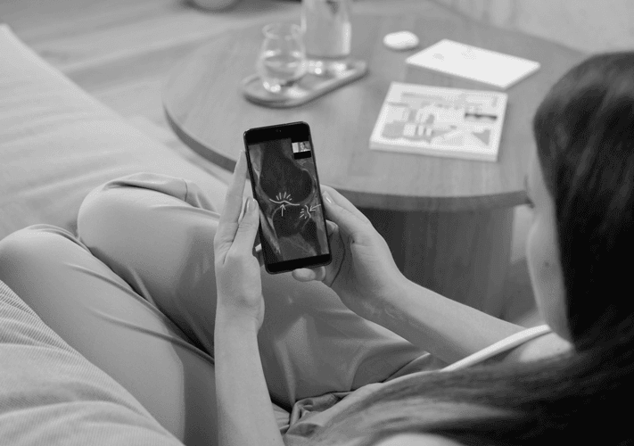
Personal explanation via video call
You will receive my explanation in a personal conversation via video call – uncomplicated, discreet and understandable.
Second opinion from a specialist - independent and online
Doubts about a treatment recommendation? Get a second opinion – independent and uncomplicated.
As a radiologist, I have the advantage of being in a neutral position, which means that I have no interest in suggesting one treatment option or another to you. Your health takes center stage.
How does it work online?
You make an appointment online. You then send us your MRI images electronically. A video call will then take place at the agreed time to discuss the MR images.
MRI shows nothing - but still pain?
Some complaints have functional rather than morphological causes that are not visible on an MRI. I will help you to understand this. However, it is also possible that the cause of your pain is not shown at all because it is referred pain. Another possibility is that the MRI examination needs to be extended with additional sequences to show the cause of your pain or that another examination modality, such as computer tomography, is necessary to identify the cause of your pain. For all these possibilities, I will explain to you what needs to be done step by step.
These causes often remain unrecognized – I can bring structure to your complaints.
How I can help you to classify this correctly
Thanks to many years of experience, I can recognize correlations that go beyond the mere image findings or I can show you which specialist field or professional group should be consulted further.
MRI / Radiology



Useful information

Meniscal tear on MRI
Meniscal tear
A meniscus tear is a common type of knee injury. The meniscus is a crescent-shaped cartilage in the knee joint that distributes weight in the joint and absorbs shocks.
In younger people, meniscus tears often occur as a result of sports injuries in which the knee is bent and twisted. Orthopaedic/trauma surgeons then often offer various operations to treat this: minimally invasive meniscus surgery including meniscus suturing, removal of the damaged tissue and – if possible – meniscus transplantation. In addition to meniscus repair, orthopaedic/trauma surgeons may also recommend surgery to realign the leg bones in patients with bow legs or knock knees in order to relieve pressure on the knee. Post-operative rehabilitation is an important part of helping patients return to their sporting and everyday activities.
In older people, meniscus injuries are usually caused by wear and tear, as the cartilage degrades with age and becomes more susceptible to tears. These patients often benefit from non-surgical measures such as physiotherapy and injections to help them resume their daily activities.
Surgery for meniscus tears
The type of surgery your doctor recommends usually depends on where the meniscus is torn, how severe the tear is and what it looks like – all this information is assessed together with a physical examination, X-rays and MRI images.
As a rule, attempts are also made to preserve the meniscus as much as possible during surgical procedures, as it plays an important role in shock absorption and weight distribution in the knee and in the mobility of the knee joint.
Operation with meniscus suture
MRI images are used to determine the blood supply in the area of the meniscus tear and the type of tear in order to decide whether meniscus suturing is even possible. Meniscus suturing has the great advantage that no meniscus tissue usually has to be removed, so that the meniscus is still available for shock absorption and weight distribution in the knee joint and can thus prevent premature wear of the cartilage layers on femur und tibia.
To suture the meniscus, surgeons perform arthroscopic surgery, in which only two to three small incisions need to be made in the knee instead of one large open incision. The surgeon inserts a tiny camera through one of these incisions; suture instruments are then inserted through the other small incisions in order to suture the meniscus tear.
This minimally invasive technique enables faster healing and recovery than a larger incision in so-called “open” operations and usually takes around 30 – 60 minutes.
After meniscus suture surgery, it is recommended to walk with crutches for up to six weeks to take the strain off the knee. Physiotherapy treatments help patients regain most of the functions of their knee within two to three months, although a full recovery can take up to six months.
Partial meniscectomy surgery
In a partial meniscectomy, damaged meniscus tissue is removed. Orthopaedic/trauma surgeons recommend this operation if the MRI images indicate that the damaged area of the meniscus is poorly supplied with blood or that the tear pattern is irreparable. A tear in the meniscus can lead to a state of irritation or inflammation in the knee joint due to partially displaced parts or irregular edges on the surface of the meniscus, causing pain.
Partial meniscectomies are usually performed arthroscopically (see meniscus suture surgery above). The surgeon removes the part of the meniscus that is irritating the knee joint and preserves as much of the meniscus as possible; the surface of the remaining meniscus is smoothed at the same time. The entire procedure takes about 30 minutes.
After partial meniscectomy surgery, you may need to use a cane or crutches for a week. Full knee function is usually restored 3 to 6 weeks after the operation with an appropriate rehabilitation program.
Meniscus transplant surgery
The surgeon may recommend a meniscus transplant if the MRI images show that you have a large or severe tear of the meniscus that requires the removal of most of the meniscus. Without a meniscus, you have an increased risk of developing osteoarthritis in your knee. If you already have osteoarthritis in your knee, your doctor may discuss the possibility of a total joint replacement with you.
In a meniscus transplant, the surgeon removes the rest of the damaged meniscus from the joint and replaces it with a meniscus of a similar size from a donor.
After a meniscus transplant operation, a splint must be worn and you may only walk with crutches for 4 to 6 weeks. Physical therapy can help you to resume everyday activities after just 2 to 3 months, while it can take 6 months to a year before you can resume sporting activities.
Meniscus surgery in combination with osteotomy
X-ray examinations can show that there is a meniscus tear together with a misalignment of the leg bones. In this case, your doctor may recommend an operation to realign the leg bones, known as an osteotomy, in addition to the actual meniscus operation.
During an osteotomy, your surgeon makes an incision in the knee at the point where the bone needs to be corrected. Your surgeon then realigns the leg bones and the knee joint by removing a small wedge of bone or inserting an implant at the end of the femur or tibia. This procedure usually takes about one to two hours.
After meniscus surgery in combination with an osteotomy, the knee should be supported by crutches for up to 6 weeks; this is followed by an individual physiotherapy program lasting several months to support recovery.
Stem cell therapy
Stem cells are cells that can transform into different types of tissue in the body. They are obtained from the patient’s blood, fatty tissue or bone marrow and can then be used to support the healing of the meniscus tissue.
Non-surgical treatments for meniscus tears
In older people, the injury may be the result of degenerative meniscus tissue due to general wear and tear, which your doctor can determine with the help of X-rays and MRI images. Treatment of these tears usually begins with reduced weight bearing, ice, compression, elevation, pain-relieving medication, physiotherapy, pain injections and, if necessary, stem cell therapy.
Rest, ice, compression and elevation
To treat a meniscus tear, your doctor may recommend rest, ice, compression and elevation in the acute phase.
Resting the knee can help to relieve symptoms. Your doctor may suggest using a cane for a few weeks to take pressure off your knee and avoid physical activities that may have contributed to the injury.
In the first few days after a meniscus tear, applying ice to the painful area and regularly elevating the knee can reduce the swelling. Wearing a compression bandage can also reduce swelling in some cases.
Pain-relieving medication
Anti-inflammatory medication can help to relieve the inflammation and pain caused by a meniscus tear.
Physical therapy
Once the inflammation in the joint has subsided and patients can stand and walk without major pain, physiotherapy is often recommended to restore strength and mobility in the injured knee. Physiotherapists can draw up an individual treatment plan to enable patients to resume everyday activities.
Strengthening the thigh and leg muscles and stretching the knee, thigh and leg can help to restore the full range of motion of the knee. Low-impact exercises such as stationary cycling can reduce pain, improve mobility and restore function to the area around the meniscus tear. As the muscles in the knee area regain strength, the physiotherapist will guide the patient to gradually return to more strenuous exercise.
Infiltrations
During infiltrations, anti-inflammatory medication is injected into the knee joint with a thin needle, which can relieve knee pain. This is not expected to heal the meniscus tear, but the pain cycle can be interrupted and swelling and discomfort can be reduced. Sometimes long-lasting pain relief can also be achieved.
You can return home or to work immediately after such an infiltration. The advantages and disadvantages of this therapy must be discussed individually with the patient.
Stem cell therapy
Stem cells are cells that can transform into different types of tissue in the body. They are obtained from the patient’s blood, fatty tissue or bone marrow and can then be used to support the healing of the meniscus tissue.

Disc herniation with compression of the nerve root on MRI
Back pain
Back pain is one of the most common types of pain. The causes are varied and range from injuries to the spine, wear and tear on the intervertebral discs, pinched nerves, muscle tension and congenital misalignments to overloading, inflammation, infections, tumors or psychosomatic influences.
Treatment options for back pain
Doctors support many sufferers with targeted measures such as movement adjustments, painkillers, infiltrations, physiotherapy, minimally invasive techniques or surgical interventions.
Use of medication
Medication is usually prescribed as a first measure against back pain. Even if they do not directly eliminate the causes, they can temporarily alleviate the discomfort and enable the patient to start physiotherapy or an exercise program – which in turn can contribute to long-term improvement.
Certain diseases such as bulging discs or degenerative changes in the intervertebral discs often impair nerve function. If, for example, a nerve root is impaired by a slipped disc or a bone spur, the nerve signals may be interrupted, resulting in painful muscle spasms. In such cases, muscle relaxants can help to relieve these spasms and reduce pain.
Infiltrations for back pain
If pain medication alone does not help sufficiently and there is a pinched nerve or a bulging disc, infiltration may be considered. This involves injecting an anti-inflammatory medication specifically into the vicinity of the affected nerve or into the fluid-filled space around the spinal cord. In order to ensure precise placement, the procedure is carried out using imaging – such as X-ray, ultrasound or CT – and usually takes less than 30 minutes.
Relief can last from a few days to a year or longer – especially if the pain cycle is broken. Infiltrations are often particularly effective in combination with physiotherapy, as they make it possible to build up muscles again without pain.
Physiotherapy
Physiotherapy plays a central role in the treatment of back pain and can help to prevent further damage to the spine. The therapy plans are individually adapted to the patient’s everyday life, pain region and personal goals.
Even if medication alleviates symptoms, the targeted development of core muscles remains an essential part of therapy. Weak abdominal, thigh or buttock muscles put additional strain on the spine, which can lead to instability or a worsening of existing problems. Physiotherapists use targeted exercises to strengthen and improve posture in order to relieve nerves and reduce pain.
In addition, physiotherapy can include complementary measures such as stretching exercises, heat or cold therapies and acupuncture to promote general well-being during muscle building.
Excess weight is an additional risk factor: excess abdominal fat in particular can pull the spine forward and lead to incorrect loading. In the case of pre-existing problems such as herniated discs, excess weight increases the pressure on the spine and therefore also the risk of nerve damage. Although losing weight through exercise is often more difficult, it can help to reduce pain in the long term.
Surgical interventions
If paralysis worsens or if conservative treatments (medication, physiotherapy, injections) do not bring sufficient improvement, surgery may be necessary.
One example is spinal fusion, for example when a vertebra slips out of place and thus narrows the spinal canal or presses on a nerve. The misalignment is corrected and disruptive tissue such as disc material, bone spurs or thickened ligaments are removed. The aim is to create space again for the spinal cord and nerve roots.
Stabilization is achieved using screws, rods or a bone graft. Over time, the connected vertebrae fuse together to form a stable bone block. A possible disadvantage of this technique is the additional strain on neighboring segments, which can lead to complaints later on.
After the operation, patients initially spend several hours in the recovery room. A hospital stay of a few days is common. Physiotherapists help patients get up for the first time and give them exercises to build up their muscles. Initially, walking aids may be required, but independent mobility can usually be regained after a few weeks. In some cases, it is necessary to wear a back support (orthosis) – especially after major surgery.
Painkillers are still required after discharge. The exact duration of the medication and the healing time depend on individual factors such as age, duration of illness and previous illness. Full recovery can take six to twelve months, in complex cases even longer. During this time, regular check-ups are carried out, including diagnostic imaging (e.g. X-ray, CT or MRI) to monitor the healing process.

Complete rupture of the anterior cruciate ligament in the MRI
Tears of the anterior cruciate ligament (ACL)
A distinction is made between partial and complete tears of the anterior cruciate ligament.
Whether an operation is necessary depends on various factors. The clinical examination by the attending physician is particularly important – this checks whether the knee joint is stable or unstable. In the case of a complete tear and a lack of muscular stabilization, the clinical tests reveal an abnormally increased mobility in the joint. The patient’s age, level of activity and any concomitant injuries (e.g. other ligaments) are also taken into account in the decision. If surgery is performed, this is usually followed by a structured rehabilitation program.
Non-surgical, so-called “conservative” treatment options for cruciate ligament ruptures include joint injections, stem cell treatments, pain medication as well as physiotherapy and occupational therapy. These measures can alleviate the symptoms and improve the function of the joint.
Surgical treatment of the anterior cruciate ligament
The patient’s level of physical activity helps to decide whether reconstruction or repair of a torn anterior cruciate ligament is more suitable.
ACL reconstruction
Reconstruction is usually performed arthroscopically – this means that only two to three small incisions are required. A tiny camera with a light source is inserted into the knee through one of these incisions so that the inside of the joint can be viewed on a monitor.
In ACL reconstruction, the torn ligament is removed and replaced with a tissue graft that is fixed to the tibia and femur using screws or anchors.
To ensure optimum stability and restore the natural rotational movement of the knee, precise drill channels are created in the thigh and lower leg to position the graft correctly.
During the operation, your surgeon creates a new ACL from a piece of tissue, also known as a graft. The graft is usually taken from a healthy part of the injured knee. The use of the body’s own tissue, known as an autograft, may be recommended if you wish to return to significant physical activity or vigorous sports.
The autograft usually comes from one of three areas: the patellar tendon, which connects the kneecap to the shinbone, the quadriceps tendon, which connects the four muscles at the front of your thigh to the kneecap, or the hamstrings, which connect one of the large muscles at the back of your leg to the shinbone. The surgeon will make a 4 to 7 centimeter incision to remove the tissue graft. The incision may be slightly painful after the operation, but has no effect on your overall recovery. Only parts of the aforementioned ligaments or tendons are removed and then used as grafts.
Alternatively, the transplant can also come from a donor (foreign tissue).
After reconstruction with the patient’s own tissue, it is often necessary to wear a splint for two to six weeks; crutches are also recommended for one to four weeks.
If a donor transplant is used, the initial healing phase may be shorter, as no additional incision is required to remove the transplant.
As a rule, exercise therapy begins immediately after the operation, and a structured physiotherapy program is usually introduced within the first week.
ACL repair surgery
Depending on the MRI findings, repair of the torn ligament may also be an option – especially if the tear is very close to the attachment or origin of the ligament on the bone.
This technique involves reattaching the original ligament to the bone.
The advantages of this method are a shorter recovery time and the ability to return to sporting activities more quickly. In addition, there is no need to remove tissue from the knee, which can contribute to a faster recovery.

Frequently asked questions about MRI evaluation online (FAQ)
How can I have my MRI findings explained?
First make an appointment online. Then send us your MRI images electronically. We will then discuss these personally via video call at the scheduled appointment.
Is an MRI second opinion online useful?
Yes, especially if there is uncertainty about a diagnosis or treatment recommendation.
What does the explanation of my MRI findings cost?
You will be informed of the costs transparently when you book the appointment. No hidden fees.

