
Get a picture of your pain for yourself
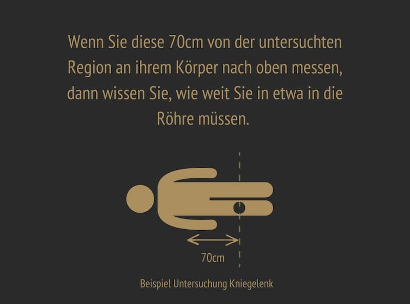
MRI arm how far into the scanner etc.
MRI arm how far into the scanner – your questions answered clearly
Many patients ask themselves: MRI arm how far into the scanner? The examination of the wrist, hand and elbow joint is usually carried out in the so-called “Superman position”, in which patients lie on their belly and stretch the arm to be examined forwards. For this reason, the head is usually still in the tube during these examinations; however, it depends on the patient’s height and the model of the MRI scanner. The procedure is painless and usually takes a maximum of 20 minutes.

MRI for elbow pain, wrist pain or thumb pain
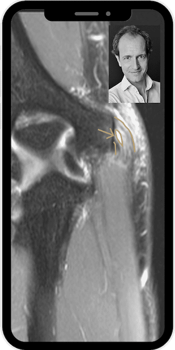
Elbow pain and MRI
Elbow pain can have many causes – from tennis elbow to osteoarthritis. An MRI can precisely visualize soft tissue and bone structures. This makes it possible to see whether a tendon insertion is inflamed, whether it is a torn tendon or whether there are signs of an irritation. Damage to the cartilage and bone marrow edema can also be shown on the MRI images.
Wrist pain - precise diagnostics with MRI
In case of wrist pain, especially pain on the outside of the wrist, an MRI often provides the most accurate diagnosis. It shows fine ligament injuries, irritations or the onset of arthrosis. The so-called discus triangularis, a washer-like soft tissue structure on the outer wrist, can also be assessed using MRI; there may be signs of wear and/or tears. The most common cause of pain in the wrist is tendinitis, but wear and tear of the joints (osteoarthritis) and pathological changes in the nerves running through the wrist (carpal tunnel syndrome) can also be diagnosed using MRI.
Pain in the little finger side of the hand - what's behind it?
Pain on the little finger side of the hand can be caused by overloading, nerve irritation or ligament problems. The MRI can detect even the smallest changes, particularly in the disc-like soft tissue structure (discus triangularis) located here. Tendon sheath inflammation can also be identified as a cause of pain using MRI.
Pain in the thumb - important structures in view
Pain in the thumb often occurs with osteoarthritis or overloading of the thumb saddle joints. MRI can be used to visualize cartilage damage, inflammation or ligament instability. MRI also has the advantage of being able to directly visualize activations / irritations of existing osteoarthritis / wear and tear of the joints. Injuries to ligaments and tendons, e.g. after skiing accidents, are also clarified using MRI.

Easy-to-understand explanation of your MRI images
I explain MRI images / MRI examinations in an easy-to-understand and uncomplicated way – whether it’s elbow pain, pain in the thumb or the question of how far the MRI arm goes into the tube.
Uncomplicated MRI second opinion online - personal and trustworthy
You can make an appointment for a personal consultation with Dr. Friedrich for an MRI second opinion online from the comfort of your own home and hold the personal consultation via video call.
Trust through specialization
Many patients, including numerous top national and international athletes, trust Dr. Friedrich due to his specialization in MRI of the joints and MRI of the spine and his resulting experience in this field.
FAQ – frequently asked questions about the MRI arm
Do I have to go all the way into the scanner for an MRI of the arm?
No – but usually the head is also at least partially in the MR scanner. The scanner focuses specifically on the region to be examined.
How long does the MRI of the arm take?
The examination takes a maximum of 20 minutes, depending on the question.
What does the evaluation and discussion of my MRI images cost?
You will receive specialist medical consultation at a fair fixed price – neutral and independent. You will find out the exact costs in advance when you make your appointment.

Upload your MRI images and receive a second opinion
Take the opportunity to upload your MRI images and get an independent second opinion from an experienced radiologist and professor of musculoskeletal radiology specializing in joints and spine. Understand your findings better and make informed decisions about your health.

