
Get a picture of your pain for yourself
MRI cervical spine & MRI sacroiliac joints & MRI lumbar spine Explanation
Your MRI of the cervical spine, lumbar spine or sacroiliac joints – explained clear and independently.
Whether neck, back or low back pain: MRI examinations of the spine provide valuable information – but often in technical language that is incomprehensible to laypeople. I will explain your MRI images of the cervical spine (cervical spine), lumbar spine (lumbar spine) or sacroiliac joints (SIJ) to you – independently, straightforwardly and in a way that is easy to understand.
MRI lumbar spine (l-spine) – clearly explained
Lumbar spine MR images Explanation
MRI of the lumbar spine: provides information about the intervertebral discs, vertebral bodies (lumbar vertebral body), facet joints, the spinal cord and the surrounding tissue. Inflammation, herniated discs or degenerative changes such as slippage of the vertebral body due to wear and tear (spondylolisthesis, e.g. Meyerding grade 1) can also be detected. With an MRI lumbar spine image explanation, you can get an idea of your symptoms yourself, which is valuable for upcoming treatment decisions and compliance with the chosen treatment.
FAQ about MRI lumbar spine
MRI lumbar spine how far into the scanner do I have to go?


Do I have to go all the way into the tube for a lumbar spine MRI? In most cases, a partial scan is sufficient – i.e. you only have to lie with your lower body in the tube for a lumbar spine MRI. During an MRI examination, the body region to be examined must be positioned in the middle of the MR scanner. Most modern MRI machines are approx. 140 cm wide, so that the center of the MR scanner is around 70 cm. With this knowledge, it is easy to estimate before the examination how far into the tube you will have to go and whether your head will be outside the tube or not. The diagrams below can help you to visualize this. Many modern devices are spacious, i.e. they have a larger tube diameter, which makes them particularly suitable for patients with claustrophobia.
MRI lumbar spine procedure
Arrive on time for your MRI examination at the agreed appointment. You should first report to the counter and register. There you will be given important information about the examination and usually also information material, which you should then read through at your leisure and, if necessary, carefully complete the enclosed questionnaires. As soon as you are called to the waiting area, you will be taken to the changing room, where you will usually be informed about the examination. You will also be asked to clear the area to be examined and to remove all metal objects, electronic devices or other objects sensitive to electromagnetic radiation; if you have any questions about this, please do not hesitate to ask the staff at the respective institute. You will then be placed in the MR scanner. The examination itself is straightforward: you will lie in the tube for about 10 to 20 minutes while the images are taken. The duration depends on the question being asked, the image quality and the device. It is important that you lie very still during the MR examination and do not move, as otherwise the images will be blurred and less easy to assess. It should be noted that, for example, movements of the head or toes have a negative effect on the image quality during an MRI of the lumbar spine, because movements of more distant body regions can also have a negative effect on the image quality.
MRI lumbar spine which organs can be seen?
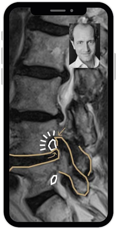
MRI lumbar spine explanation - typical findings
- MRI lumbar spine disc herniation: The German term “Bandscheibenvorfall” was replaced in international specialist circles in 2011 by terms such as herniation, bulging, protrusion or extrusion of the disc. All of these terms describe protrusions of the intervertebral disc, which can take different shapes; the important thing is whether the bulging disc is compressing a nerve or the spinal cord and thus causing discomfort. An MRI of the lumbar spine can show this very well.
- Lumbar spine degeneration (e.g. “degenerative changes”, “osteochondrosis”): Wear and tear of the intervertebral discs, vertebrae, vertebral joints, ligaments, joint capsules and muscles in the lumbar spine are referred to as lumbar spine degeneration. Lumbar spine degeneration increases with age and can lead to pain.
- Sciatica irritation – what is the sciatica trying to tell me? The sciatica is a large nerve that supplies both the legs and feet. Irritation of the sciatic nerve can cause pain and can be visualized in the MRI of the lumbar spine.
- Malalignments: Lordosis Kyphosis; kyphosis and lordosis are curvatures of the spine. A normal thoracic spine has a certain degree of kyphosis and a normal lumbar spine has a certain degree of lordosis, but too much kyphosis and lordosis or kyphosis of the lumbar spine and lordosis of the thoracic spine can also be pathological and associated with pain. Although kyphosis and lordosis are visible in the MRI, they must be confirmed by means of a standing image (usually X-ray) in order to make a correct diagnosis.
- MRI lumbar spine inflammation: Inflammation of the intervertebral discs and the adjacent vertebrae and soft tissue can cause pain. An MRI of the lumbar spine can identify such inflammation as the cause of pain.
Experienced specialist expertise
Benefit from the many years of experience of a specialist in MRI of the joints and spine, who will analyze your MR images precisely and explain them in a way that is easy to understand.
Independent assessment
Our assessments are free of any vested interest in one or the other of the further treatments, so that you receive a completely independent assessment.
Personal Consultation
Receive a personalized explanation of your findings from Dr. Friedrich, who will address your specific questions and concerns.
MRI cervical spine (c-spine) – what do the findings mean?
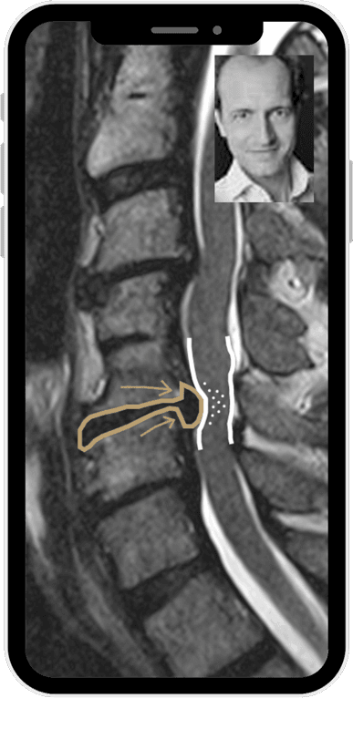
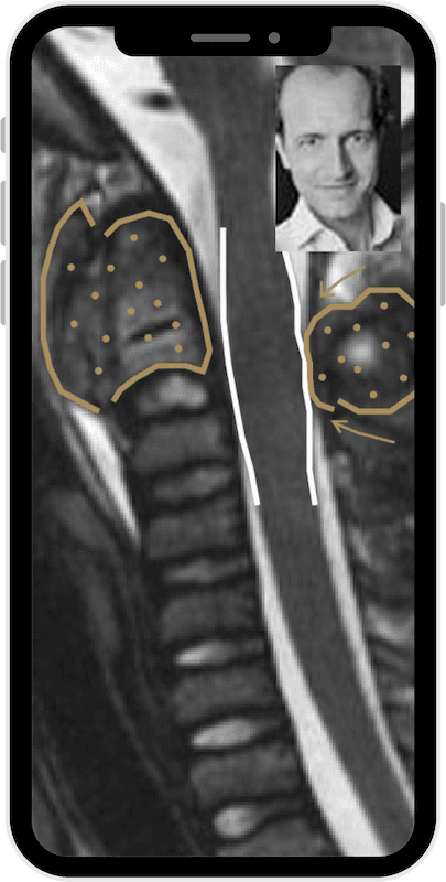
When is an MRI of the cervical spine useful?
Some diagnoses in the MRI cervical spine
- Herniated cervical spine disc
- Spinal canal stenosis cervical spine, neuroforaminal stenosis cervical spine
- MRI cervical spine tumor (e.g. metastases or benign masses)
- Wear and tear of the small vertebral joints – can cause neck and shoulder pain.
What can we really read from the MRI?
I will explain the medical terms to you and show you whether and which treatment makes sense – without any interest in therapies or operations.
evaluate MRI SIJ (sacroiliac joint) and sacral bone
When does an MRI of the SI joint make sense
Deep-seated low back pain that occurs on one or both sides is typical. SI joint complaints are often overlooked or dismissed as “non-specific”.
OA of SI joints MRI
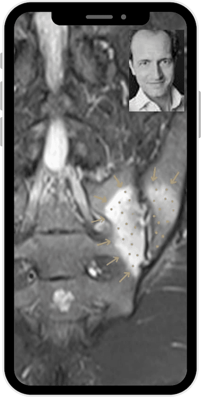
Joint wear at the sacroiliac joint (SI joint) can be visualized well in an MRI. However, I will also explain to you whether other causes of pain such as inflammatory changes (sacroiliitis) can be seen at your SI joint.
What does an MRI of the sacrum / SI joint show?
- SI joint osteoarthritis or inflammation
- Broken bones (fractures) or bone bruises
- Tumors
- Anatomical variants such as lumbosacral transitional vertebrae with or without an assimilation joint.
FAQ – frequently asked questions about MRI of the spine
Here you will find answers to frequently asked questions about MRI images of the spine.
MRI lumbar spine duration?
An MRI of the lumbar spine usually takes between 15 and 25 minutes, depending on the problem and the requested image quality.
MRI thoracic spine duration?
An MRI of the thoracic spine usually takes between 15 and 25 minutes, depending on the problem and the requested image quality.
What does the term Meyerding grade 1 mean?
Meyerding grade 1 describes a slight displacement of a vertebra, known as spondylolisthesis, which usually does not require immediate surgery.
What is an SI joint block?
An SI joint block is a temporary restriction of movement in the sacroiliac joint that can cause pain in the lower back.
Can an MRI show whether a vertebra is ‘dislocated’?
Symptoms of a dislocated vertebra: pain and restricted movement. Imaging of a dislocated vertebra: the classic diagnosis of a dislocated vertebra is a manual medical or chiropractic diagnosis that is made without imaging. However, an MRI can very well show other important causes of pain and restricted movement, such as wear and tear, inflammatory changes or tumors, which should often be ruled out beforehand.
What information does an MRI of the spine provide?
An MRI of the spine can provide detailed information about the intervertebral discs, vertebral bodies and surrounding soft tissue in order to diagnose diseases or injuries.
How do I prepare for an MRI scan?
Wear comfortable clothing without metal parts and inform the medical staff of any implants or health concerns.
Upload your MRI images and receive a second opinion
Take the opportunity to upload your MRI images and get an independent second opinion from an experienced radiologist and professor of musculoskeletal radiology specializing in joints and spine. Understand your findings better and make informed decisions about your health.

