
Get a picture of your pain for yourself
MRI translation of reports for laypersons
Understand your MRI images and MRI results better
MRI images provide impressive insights into the body – whether head, spine or full-body images. However, the medical language often remains incomprehensible. Dr. Friedrich therefore helps you with an MRI report translation for laypersons and explains your MRI results independently, comprehensibly and at eye level.
MRI images – what can be seen?
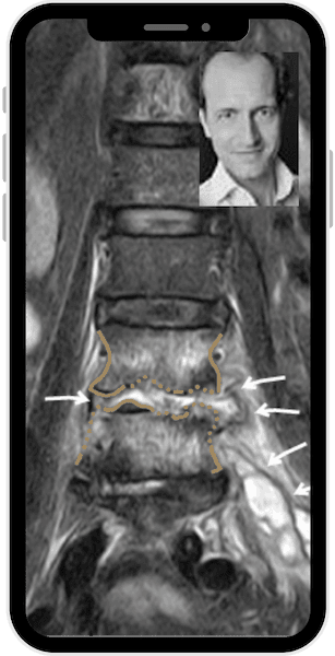
What does inflammation look like in an MRI?
Many patients are diagnosed with inflammation of the musculoskeletal system after their MRI scan, but they then ask themselves what inflammation looks like on an MRI scan. In the case of inflammation, the MRI device recognizes that more fluid is stored in the tissue – this fluid can be easily depicted in the MRI because it stands out clearly from the normal, non-fluid-impregnated tissue. This explanation is of course greatly simplified, as it is not just simple fluid, but a large number of inflammatory cells and inflammatory tissue. It is important to differentiate between acute and chronic inflammation and irritation – this is where MRI provides valuable information.
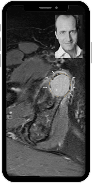
What do metastases look like on MRI?
Patients with a known tumor disease often wonder what metastases look like on MRI. Metastases often appear in the MRI as roundish signal changes – distinguishing them from harmless lesions requires experience. They can occur in the soft tissue or in the bone and in both cases can be visualized very well using MRI.
Special questions about MRI
Claustrophobia open MRI head - does it make sense?
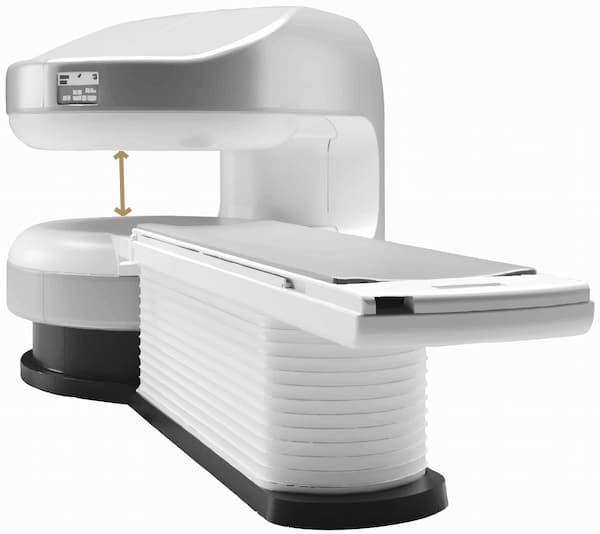
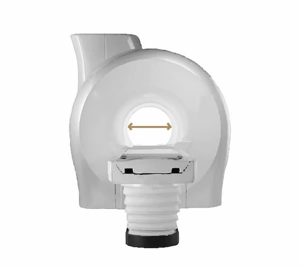
Open MRI scanners offer significantly more space for patients with claustrophobia. From a physical point of view, however, it is clear that the more closed the system, the more homogeneous the magnetic field and the more homogeneous the magnetic field, the better the image quality. However, there are now also closed MRI scanners that have a larger internal diameter than before (“large MRI”), which is very helpful for many patients with claustrophobia and still provides very good image quality. Dr. Friedrich can then explain the MRI findings to you and explain the MRI results and MRI images.
Large MRI or normal MRI - what is the difference?
MRI tattoos - is there a risk?
The ink in the tattoos may contain metal particles that react with the magnetic field. Reactions range from a slight tingling sensation, skin irritation, burning or a feeling of heat to slight burns. For this reason, you should wait at least 6 weeks before undergoing an MRI, especially after a fresh tattoo. If you feel any of these reactions in the area of your tattoo during the MRI examination, please press the emergency ball around it and inform the MRI team examining you.
MRI side effects - what is possible?
As a rule, MRI is very well tolerated. Rare side effects may occur, e.g. due to contrast agents (e.g. allergic reactions). It is important that people with pacemakers and other metallic implants in their bodies are not examined by MRI or that it is clarified in advance whether the respective implant is suitable for MRI or not. In order to be able to clarify this carefully, please disclose all implants (including implant passports) when making the appointment for the MRI.
Whole-body MRI - when is it useful?
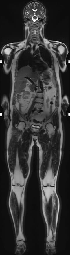
A whole-body MRI can be useful for certain cancers or as a preventive examination.
The advantages of whole-body MRI are that, unlike CT or X-ray, the examination does not require X-rays, making it suitable for repeated check-ups and preventive examinations. Whole-body MRI provides a good overview of important organ systems in the body and, like all MRI examinations, has a high soft tissue contrast. The disadvantages of whole-body MRI are the limited image quality (resolution) compared to the examination of individual parts of the body, a relatively long examination time and the fact that some organ systems cannot be adequately assessed, such as the lungs and gastrointestinal tract, but also smaller joints and vessels.
Dr. Friedrich helps you to understand MRI findings from such examinations.
Difference between CT and MRI or X-ray?
MRI or X-ray - when does which make sense?
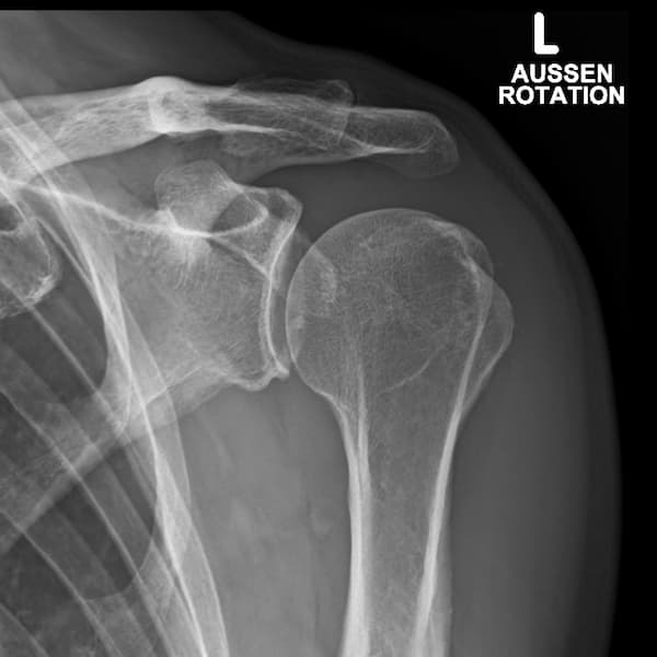
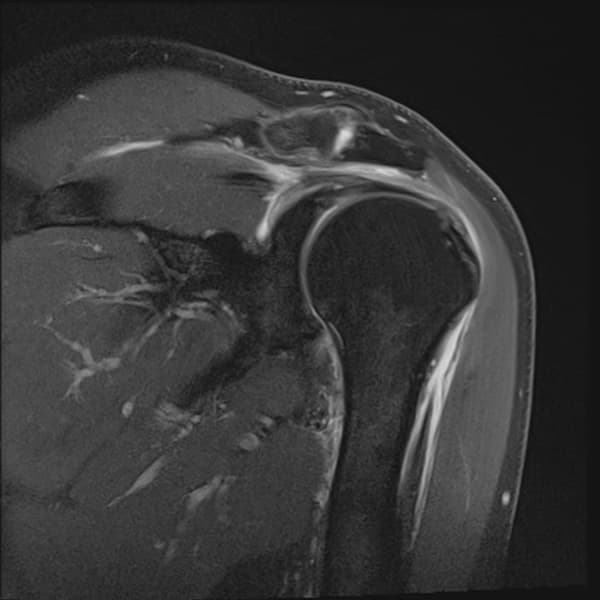
Many patients ask themselves: MRI or X-ray – which examination is better for me? There is no general answer to this question. It depends on what you are looking for and what exactly you want to see. X-rays show bony structures and calcifications well, while MRI has significantly better soft tissue contrast, can also show changes in the bone marrow earlier and more clearly and is a cross-sectional imaging procedure, meaning that structures are not superimposed and a more precise assessment of the structures is possible. Another fundamental difference between MRI and X-ray is that X-ray radiation is used to generate images, whereas MRI uses magnetic fields and radio frequency pulses to generate images.
What do you see on CT?
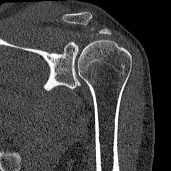
CT (computed tomography) uses X-rays and shows bones, calcifications, the lungs or bleeding very well – but less detail in soft tissue compared to MRI, so the soft tissue contrast of CT is lower than in MRI. Bone marrow edema is better visible in MRI than in CT. CT and MRI are both cross-sectional imaging techniques, so there are no overlaps that make the diagnosis more difficult. Both procedures therefore have their areas of application, their advantages and disadvantages.
Radiation exposure during MRI?
Patients often ask about the radiation exposure during MRI. MRI works with magnetic fields – electromagnetic radiation is used but not ionizing radiation or X-rays; this is what makes MRI fundamentally different from X-rays and CT.
Creatinine value MRI - why is it important?
Is it always necessary to determine the creatinine value in an MRI? When contrast media is administered, the kidney function is tested, typically via the creatinine value or the GFR. It is not necessary to determine the creatinine value for an MRI examination without contrast medium.
Your benefits with Talk2Friedrich
Independent assessment of your MRI images
Dr. Friedrich explains your MRI images in an easy-to-understand and independent manner.
Online and uncomplicated
Whether you have claustrophobia, unclear findings or specific questions, you can receive Dr. Friedrich’s independent opinion via video call, either at home or on the go.
MRI specialization
Dr. Friedrich specializes in imaging the musculoskeletal system, in particular using MRI, and is therefore happy to help you with his wealth of experience.

FAQ – frequently asked questions about MRI images
Here you will find answers to frequently asked questions about MRI images of the spine.
Is an MRI dangerous due to radiation?
What is a “large MRI”?
A large MRI is a closed MRI machine with a larger tube opening – more comfortable for patients with claustrophobia or a wider physique, while still providing very good examination quality.
How do you recognize inflammation or metastases in an MRI?
Inflammation is shown in the MRI by an increase in fluid retention in the tissue, which is clearly distinguishable from normal tissue. Metastases are shown in the MRI as roundish signal changes – precise interpretation and differentiation from harmless lesions requires appropriate specialist knowledge.

MRI second opinion online
Get an independent MRI second opinion online from Dr. Friedrich, an experienced radiologist and professor of musculoskeletal radiology specializing in joints and spine. He will help you to better understand your MRI findings so that you can make better informed decisions about your health.

